Designed with a compact and intelligent design, the 1950's shoe x-ray machine offers various modes of imaging such as full body scans and localized scans. The device supports digital viewing stations where the images can be viewed remotely by radiologists. The 1950's shoe x-ray machine assists in improved diagnostic operations by providing easy-to-handle mechanisms and sound imaging consistency.

In the hospital and clinic setting, the 1950's shoe x-ray machine is utilized for chest imaging, exposing respiratory and cardiovascular pathologies. It is widely employed to monitor pneumonia, tuberculosis, and cardiac enlargement. The 1950's shoe x-ray machine is also important in dental and maxillofacial examinations, providing precise visual markers in treatment planning.
Future versions of the 1950's shoe x-ray machine will combine energy-efficient technology with high-resolution imaging. Predictive analytics integration will enable early disease detection and personalized screening. Global telemedicine networks will also be enabled by the 1950's shoe x-ray machine, extending access to diagnosis in underserved populations.

The 1950's shoe x-ray machine needs regular maintenance to function at its best. Technicians need to regularly inspect exposure controls, cooling systems, and image sensors. The 1950's shoe x-ray machine has to be run within prescribed usage boundaries, and annual recalibration needs to be planned to maintain radiation accuracy as well as uniform imaging quality.
In today's healthcare system, the 1950's shoe x-ray machine continues to be an integral part of diagnostic imaging. The 1950's shoe x-ray machine provides precise visual data that helps in disease detection and assessment of an injury. The 1950's shoe x-ray machine has digital sensors and the capability to improve images. The 1950's shoe x-ray machine helps in quick and effective medical imaging.
Q: What makes an x-ray machine different from a CT scanner? A: An x-ray machine captures a single 2D image, while a CT scanner takes multiple x-rays from different angles to create 3D cross-sectional views. Q: How is image quality measured in an x-ray machine? A: Image quality depends on factors like contrast, resolution, and exposure settings, which are adjusted based on the target area being examined. Q: What power supply does an x-ray machine require? A: Most x-ray machines operate on high-voltage power systems, typically between 40 to 150 kilovolts, depending on their intended use. Q: Can x-ray machines be used for dental imaging? A: Yes, specialized dental x-ray machines provide detailed images of teeth, jaws, and surrounding structures to support oral health assessments. Q: How does digital imaging improve x-ray efficiency? A: Digital systems allow instant image preview, faster diagnosis, and reduced need for retakes, improving workflow efficiency in clinical environments.
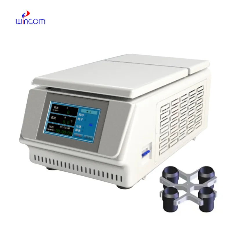
We’ve used this centrifuge for several months now, and it has performed consistently well. The speed control and balance are excellent.
This ultrasound scanner has truly improved our workflow. The image resolution and portability make it a great addition to our clinic.
To protect the privacy of our buyers, only public service email domains like Gmail, Yahoo, and MSN will be displayed. Additionally, only a limited portion of the inquiry content will be shown.
We are planning to upgrade our imaging department and would like more information on your mri machin...
I’m looking to purchase several microscopes for a research lab. Please let me know the price list ...
E-mail: [email protected]
Tel: +86-731-84176622
+86-731-84136655
Address: Rm.1507,Xinsancheng Plaza. No.58, Renmin Road(E),Changsha,Hunan,China

 af
af
 es
es
 ar
ar
 tr
tr
 sw
sw
 pt
pt
 th
th
 ur
ur
 bn
bn
 ne
ne
 vi
vi
 km
km
 lo
lo
 de
de
 ru
ru
 fi
fi
 nl
nl
 fa
fa
 fr
fr
 ko
ko