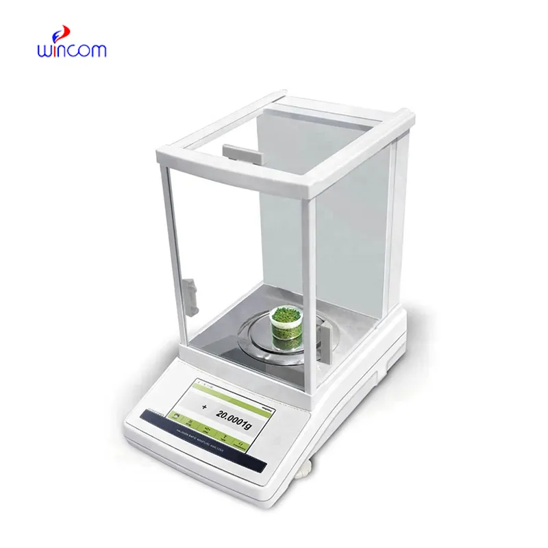
In hospital and clinical laboratories, electronic balance scale supplies exact mass measurements that are very important to experimental integrity. It allows the very accurate weighing of the reagents, chemicals, and components of the patient's sample. Technicians in the laboratory use electronic balance scale for the maintenance of reproducibility, validation of method, and monitoring of therapy. electronic balance scale assures the consistency of analytical data, so, it increases the efficiency of the hospital laboratory, makes the research more reliable, and through accurate measurement of the materials in the laboratory, it becomes part of the patient care that is of high quality.
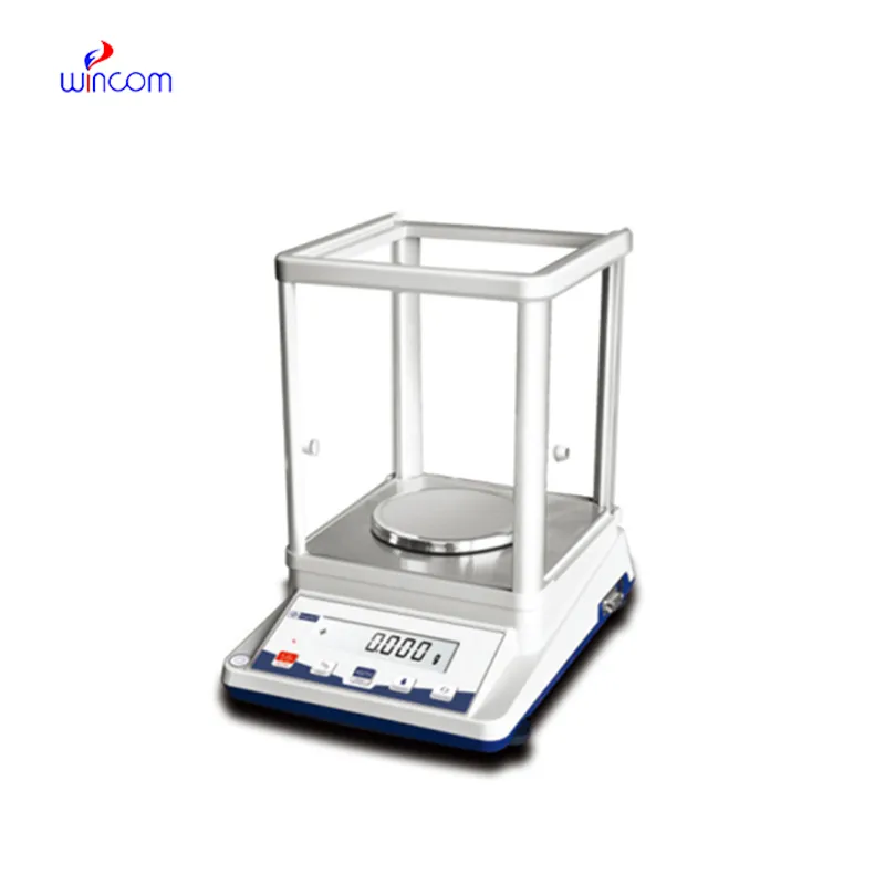
electronic balance scale is commonly used in the compounding of medicinal and chemical substances in small quantities in hospital pharmacy laboratories. Mass control with high precision is critical when dealing with active pharmaceutical ingredients in micro or milligram quantities. This application helps to prepare exact doses, internal validation, and experimental drug research. By allowing for repeatable measurements, electronic balance scale helps pharmacists and researchers in controlling formulation processes, which makes it easier to get consistency and reliability throughout hospital medication development workflows.
The ongoing evolution of regulations will see the electronic balance scale adding features for compliance support that will be more advanced. Hospital lab audits and accreditation standards will be met with the help of the documentation functions and secure data storage. This future trend will greatly improve the quality management processes in all hospitals and research labs.
electronic balance scale are used in hospital settings and are good to strict contamination control measures. So, spills on the weighing chamber must always be contained. Residue buildup that may affect sensitivity is minimized by cleaning after each weighing session. The laboratory staff can identify wear early during the inspection period scheduled for downtime, and this will support long-term measurement stability across clinical applications.
electronic balance scale plays an important role in the hospital pharmacy in the accurate formulation of medications, intravenous solutions, and compounded drugs. Even slight alterations in the weight of drugs can change the effectiveness of the drug and endanger the patient's safety. The Pharmacy Technicians rely on electronic balance scale for the correct dosing and checking of the active ingredients. The tool's accuracy guarantees the dependable preparation of drugs, the observance of the rules, and the quality control of the overall hospital pharmacy activities.
Q: What maintenance does an Analytical Balance require? A: A periodic cleaning, checking of the calibration, and also verifying the performance are all necessary. Q: Can an Analytical Balance handle continuous daily use? A: Yes, provided that the correct laboratory conditions and rules are followed. Q: Why is leveling important for an Analytical Balance? A: The accuracy and repeatability of the measurements depend on proper leveling. Q: Can Analytical Balances be connected to laboratory systems? A: Most of the models allow connectivity with laboratory information systems. Q: Are Analytical Balances sensitive to vibration? A: Yes, stable weight readings can be disturbed by vibrations.
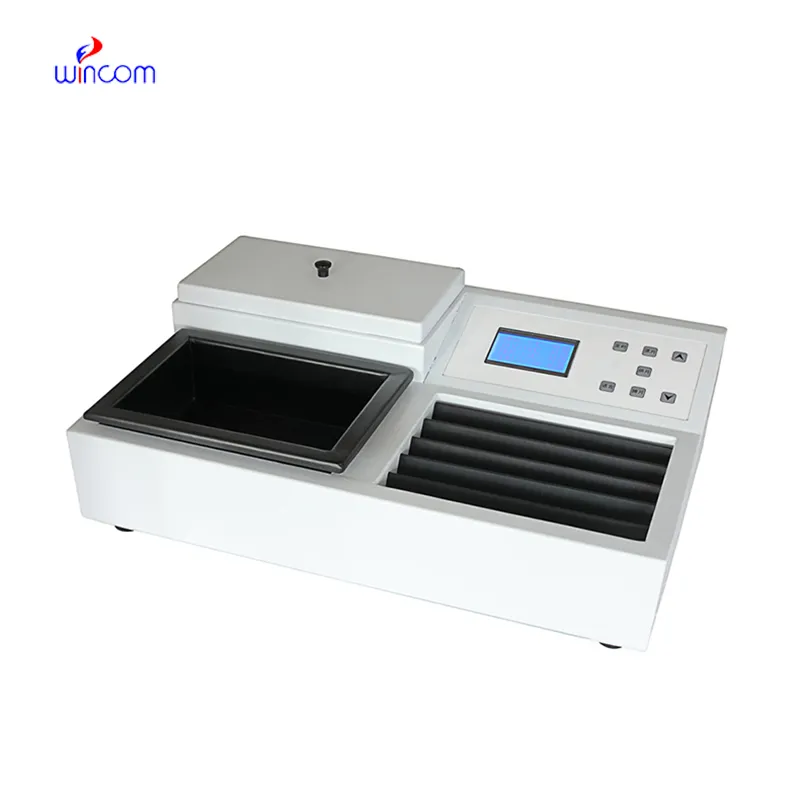
The water bath performs consistently and maintains a stable temperature even during long experiments. It’s reliable and easy to operate.
We’ve been using this mri machine for several months, and the image clarity is excellent. It’s reliable and easy for our team to operate.
To protect the privacy of our buyers, only public service email domains like Gmail, Yahoo, and MSN will be displayed. Additionally, only a limited portion of the inquiry content will be shown.
We are planning to upgrade our imaging department and would like more information on your mri machin...
Could you share the specifications and price for your hospital bed models? We’re looking for adjus...
E-mail: [email protected]
Tel: +86-731-84176622
+86-731-84136655
Address: Rm.1507,Xinsancheng Plaza. No.58, Renmin Road(E),Changsha,Hunan,China

 af
af
 es
es
 ar
ar
 tr
tr
 sw
sw
 pt
pt
 th
th
 ur
ur
 bn
bn
 ne
ne
 vi
vi
 km
km
 lo
lo
 de
de
 ru
ru
 fi
fi
 nl
nl
 fa
fa
 fr
fr
 ko
ko