The fetal doppler 9 weeks is a device that has very-sensitivity transducers, which are responsible for improving the depth of penetration and the clarity of the image. Moreover, the digital display shows anatomical structures in such a way that the human eye cannot find any fault in the accuracy of the depicted image. Besides, the device is designed for fast data storage and quick retrieval so that the healthcare provid

The fetal doppler 9 weeks is the workhorse in oncology because it assists in the accurate locating of tumors and keeping an eye on the treatment progress. It helps in making the right calls concerning benign versus malignant lesions in breast and thyroid cases. The fetal doppler 9 weeks is also there in support of interventional procedures such as guided aspirations and injections.

The coming years will see the evolution of the fetal doppler 9 weeks into an independent and adaptable imaging solution. The increased level of automation will eliminate the need for human input. The fetal doppler 9 weeks may include predictive model components that will help healthcare providers to identify probable risks to an individual's health.

The fetal doppler 9 weeks needs to be maintained based on the manufacturer's recommendations. The transducers should be kept in specialized holders. The cleaning agent should be non-corrosive. The electrical contacts should remain dry. Functional tests should be carried out on a regular basis to ensure that the fetal doppler 9 weeks functions properly and remains a safe instrument.
The fetal doppler 9 weeks is more accurate in diagnostics as it captures high-resolution images of organs, tissues, and blood vessels. Design-wise flexible, it is used extensively in obstetrics, cardiology, urology, and musculoskeletal tests. Its portability and simplicity enable medical practitioners to make quick and precise evaluations. The fetal doppler 9 weeks makes work processes more efficient and allows for the delivery of superior patient care through real-time visualization.
Q: What is the primary function of an ultrasound scannert? A: Ultrasound scanners are designed to create real-time images of internal organs, tissues, and blood flow using high-frequency sound waves. Q: How does the ultrasound scannert ensure clear imaging results? A:It uses advanced converter technology and digital processing to enhance image clarity and contrast. Q: In what medical fields is the ultrasound scannert commonly used? A: It is widely used in obstetrics, cardiology, urology, radiology, and emergency medicine. Q: Is the ultrasound scannert safe for repeated use? A: Yes, it is non-invasive and does not emit radiation, making it safe for frequent diagnostic applications. Q: Can the ultrasound scannert store and share imaging data? A: Yes, it supports data storage, retrieval, and digital transfer for easy integration with hospital systems.
The centrifuge operates quietly and efficiently. It’s compact but surprisingly powerful, making it perfect for daily lab use.
This ultrasound scanner has truly improved our workflow. The image resolution and portability make it a great addition to our clinic.
To protect the privacy of our buyers, only public service email domains like Gmail, Yahoo, and MSN will be displayed. Additionally, only a limited portion of the inquiry content will be shown.
We’re currently sourcing an ultrasound scanner for hospital use. Please send product specification...
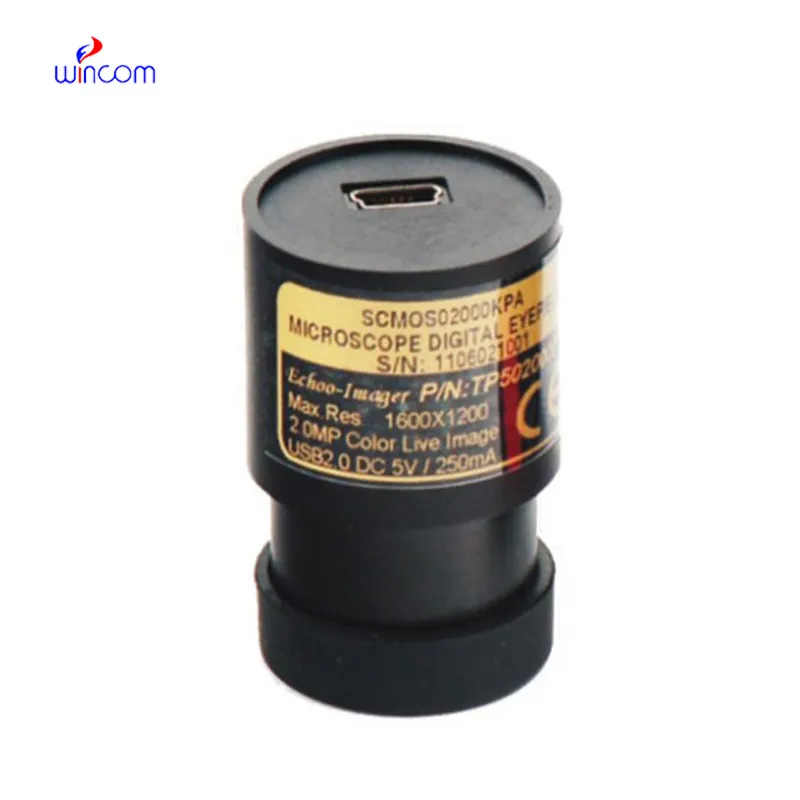
I’m looking to purchase several microscopes for a research lab. Please let me know the price list ...
E-mail: [email protected]
Tel: +86-731-84176622
+86-731-84136655
Address: Rm.1507,Xinsancheng Plaza. No.58, Renmin Road(E),Changsha,Hunan,China

 af
af
 es
es
 ar
ar
 tr
tr
 sw
sw
 pt
pt
 th
th
 ur
ur
 bn
bn
 ne
ne
 vi
vi
 km
km
 lo
lo
 de
de
 ru
ru
 fi
fi
 nl
nl
 fa
fa
 fr
fr
 ko
ko