The hand held doppler fetal monitor is a device that has very-sensitivity transducers, which are responsible for improving the depth of penetration and the clarity of the image. Moreover, the digital display shows anatomical structures in such a way that the human eye cannot find any fault in the accuracy of the depicted image. Besides, the device is designed for fast data storage and quick retrieval so that the healthcare provid

The hand held doppler fetal monitor has demonstrated its irreplaceable nature in prenatal screening, cardiovascular diagnostics, and overall health evaluations.The hand held doppler fetal monitor is a technique that evaluates organ function, reveals pathological changes, and supports medical education by providing live imaging demonstrations. The hand held doppler fetal monitor technology gives doctors the ability to perform accurate and instantaneous assessments in a variety of clinical situations.

The next-generation hand held doppler fetal monitor solutions come with better processing capabilities and intelligent algorithms that improve the clarity of images in addition to lessening reliance on operators. The aspect of augmented reality will change the world of surgical operations. The hand held doppler fetal monitor solutions will also change the face of delivering healthcare by facilitating quicker and more accurate diagnoses.

The daily upkeep of the hand held doppler fetal monitor involves cleaning, inspection, and proper storage. The removal of the gel residue from the probes should be accomplished as soon as the analysis has been carried out. The cooling vents of the device should always be unblocked. The hand held doppler fetal monitor needs annual professional servicing in order to remain accurate.
The hand held doppler fetal monitor is an essential diagnostic modality in the modern healthcare system, permitting the non-invasive imaging of internal organs and tissues. By transmitting sound waves and reading their echoes, it provides real-time data on physiology. The hand held doppler fetal monitor makes precise diagnoses feasible in all specialties, improving clinical decision-making and patient confidence.
Q: What are the main maintenance requirements for the ultrasound scannert? A: Regular cleaning, proper probe handling, and scheduled inspections help maintain optimal performance. Q: How often should the ultrasound scannert be calibrated? A: Calibration frequency depends on usage levels, but periodic professional checks are recommended. Q: Is the ultrasound scannert suitable for pediatric use? A: Yes, it provides gentle, non-invasive imaging ideal for neonatal and pediatric diagnostics. Q: Does the ultrasound scannert support wireless connectivity? A: Many models include Wi-Fi or Bluetooth features for data sharing and device integration. Q: What materials are used in the ultrasound scannert construction? A: It is built with durable medical-grade components designed to withstand continuous clinical use.
The water bath performs consistently and maintains a stable temperature even during long experiments. It’s reliable and easy to operate.
The hospital bed is well-designed and very practical. Patients find it comfortable, and nurses appreciate how simple it is to operate.
To protect the privacy of our buyers, only public service email domains like Gmail, Yahoo, and MSN will be displayed. Additionally, only a limited portion of the inquiry content will be shown.
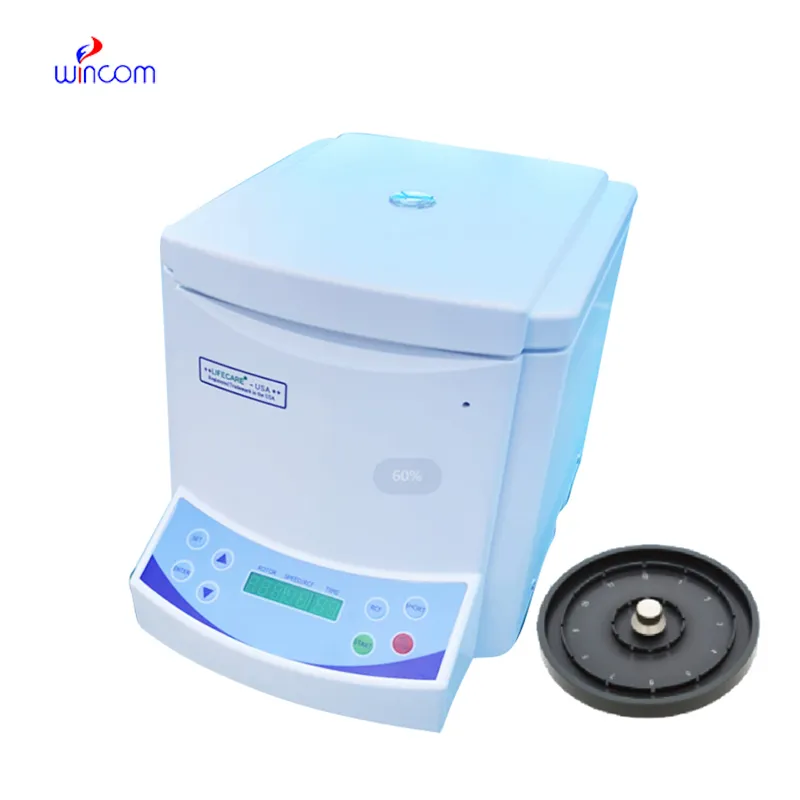
Hello, I’m interested in your centrifuge models for laboratory use. Could you please send me more ...
Hello, I’m interested in your water bath for laboratory applications. Can you confirm the temperat...
E-mail: [email protected]
Tel: +86-731-84176622
+86-731-84136655
Address: Rm.1507,Xinsancheng Plaza. No.58, Renmin Road(E),Changsha,Hunan,China

 af
af
 es
es
 ar
ar
 tr
tr
 sw
sw
 pt
pt
 th
th
 ur
ur
 bn
bn
 ne
ne
 vi
vi
 km
km
 lo
lo
 de
de
 ru
ru
 fi
fi
 nl
nl
 fa
fa
 fr
fr
 ko
ko