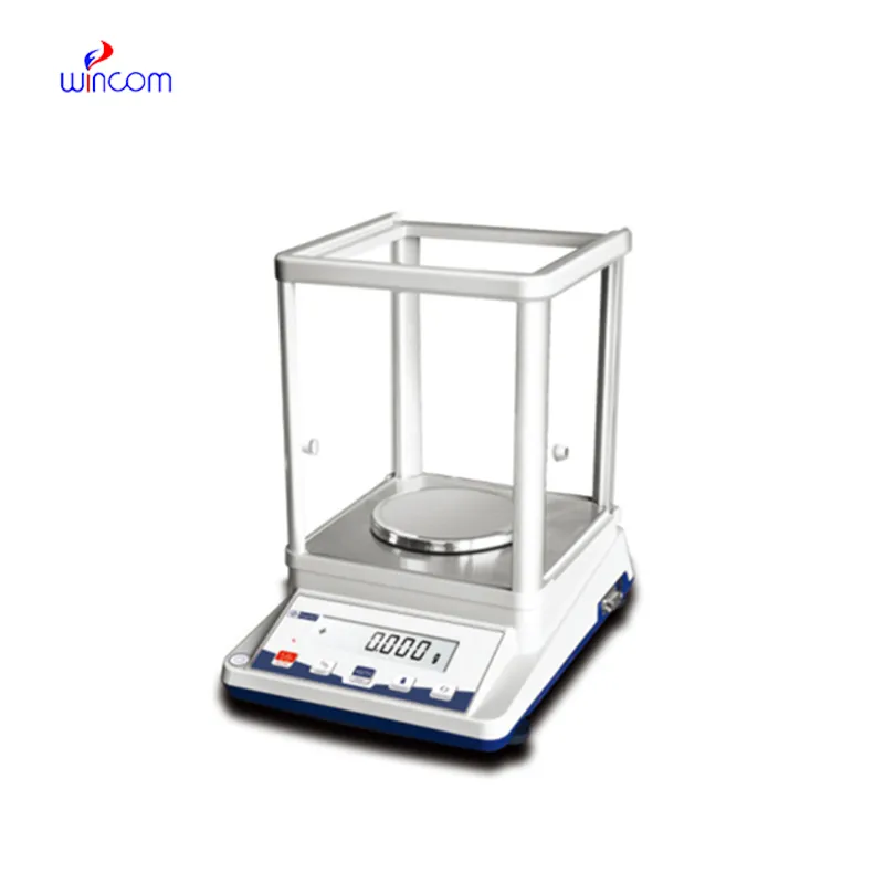
sartorius analytical balance is an extremely accurate device that is specifically designed for weighing little amounts in labs and hospital pharmacies. The instrument's sensitivity implies accurate preparation of samples for diagnostic testing, reagent making, and the production of drugs. The lab personnel consider it vital to get repeatable measurements, perform calibration checks, and validate their standards using sartorius analytical balance. The right use of sartorius analytical balance will bring about the smoothness of clinical workflows, research activities, and quality control, thus providing accuracy and reliability to all analytical processes in hospitals and laboratories.
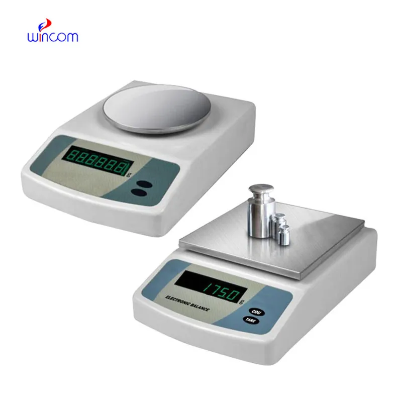
Hospitals' analytical laboratories use sartorius analytical balance during the production of internal standards for instrument testing methods. Very accurate mass input is a must to maintain uniformity over different analytical runs. This use case allows for comparison of still data, traceability, and monitoring of analytical performance over a long period. By allowing exact preparation of reference materials, sartorius analytical balance boosts measurement confidence in all phases of hospital laboratory work.
At the medical institutions that are research-driven, sartorius analytical balance will change to facilitate the analytical methods with higher sensitivity that are in the pipe. The future might bring along the possibility of ultra-low mass samples being accurately measured in molecular diagnostics and sophisticated drug research. This turning development will not only enlarge the experimental capacities of hospital-based research labs but also open new fronts in medical innovation through analytics.
For sartorius analytical balance to last long, professionals must do scheduled servicing as per the laboratory guidelines. Internal cleaning and checking of parts will help to slow down and eventually stop the performance degradation. Hospitals or labs that maintain structured service intervals not only enjoy lesser downtimes but also higher accuracy, thus they can carry on with seamless analytical operations.
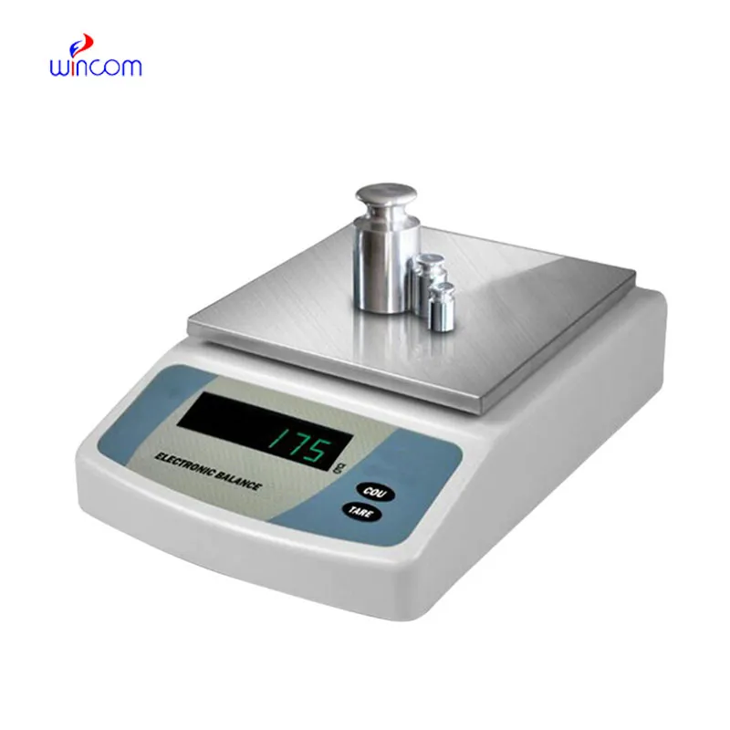
In clinical labs, sartorius analytical balance serves as the primary tool for making solutions, reagents, and calibrators that are necessary for the diagnostic tests. The accuracy of weighing determines the correctness of the results of biochemical, hematological, and immunological assays. The laboratory staff depend on sartorius analytical balance for exactness, replicability, and speed. Its function secures that the diagnostic procedures of hospitals yield valid and uniform results which in turn support patient care, treatment choices, and improved laboratory quality.
Q: What is the main purpose of an Analytical Balance? A: Its purpose is mainly to measure very tiny sample masses with the utmost precision in laboratories and hospitals. Q: What is the typical weighing range of an Analytical Balance? A: The weighing range for the majority of analytical balances is from 0 up to some grams with a resolution of micrograms or milligrams. Q: What environmental controls are necessary for an Analytical Balance's operation? A: Airflow, vibration, and temperature changes should not only be avoided but also prevented in the room where the scale is situated. Q: Is an Analytical Balance permitted in a hospital laboratory? A: Yes, it has indeed found widespread usage for the preparation of reagents, calibra¬tion, and drug development applications. Q: What should be the frequency of calibration for an Analytical Balance? A: The calibration interval is subject to the degree of use and the particular laboratory requirements.
The hospital bed is well-designed and very practical. Patients find it comfortable, and nurses appreciate how simple it is to operate.
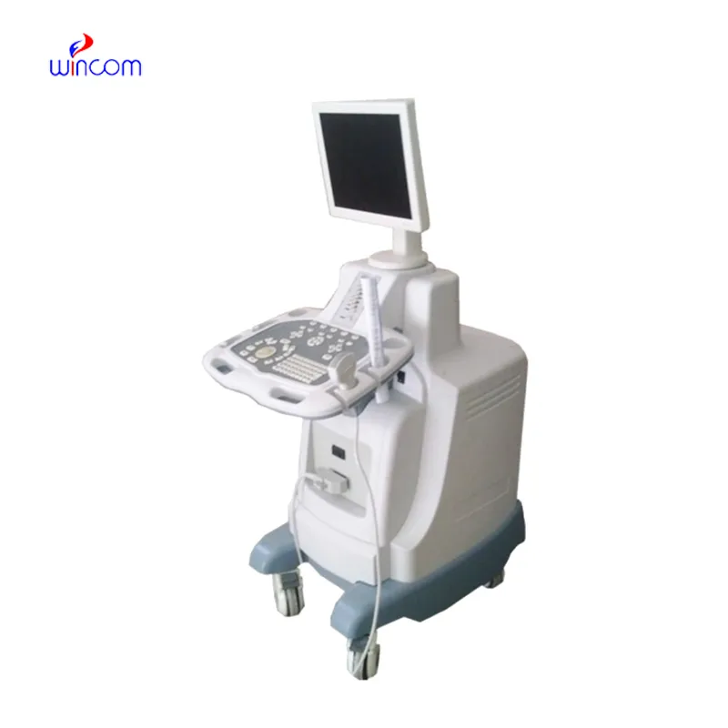
This ultrasound scanner has truly improved our workflow. The image resolution and portability make it a great addition to our clinic.
To protect the privacy of our buyers, only public service email domains like Gmail, Yahoo, and MSN will be displayed. Additionally, only a limited portion of the inquiry content will be shown.
We’re interested in your delivery bed for our maternity department. Please send detailed specifica...
We’re currently sourcing an ultrasound scanner for hospital use. Please send product specification...
E-mail: [email protected]
Tel: +86-731-84176622
+86-731-84136655
Address: Rm.1507,Xinsancheng Plaza. No.58, Renmin Road(E),Changsha,Hunan,China

 af
af
 es
es
 ar
ar
 tr
tr
 sw
sw
 pt
pt
 th
th
 ur
ur
 bn
bn
 ne
ne
 vi
vi
 km
km
 lo
lo
 de
de
 ru
ru
 fi
fi
 nl
nl
 fa
fa
 fr
fr
 ko
ko