Every feature of the sem microscope vs tem is built to provide maximum viewing accuracy and comfort to the user. The coarse and fine focus of the microscope provide control to observe large and small specimens. The sem microscope vs tem has enhanced illumination technology to give balanced light conditions to highlight color contrasts and fine details. It has also got compatibility with digital imaging software for analysis and documentation to allow researchers to store and compare results efficiently.

In medical and industrial usage, the sem microscope vs tem finds wide application. Pathologists utilize it to identify cancer cells, microbiologists to characterize bacteria, and botanists to study plant cell morphology. In electronics, the sem microscope vs tem facilitates defect analysis of printed circuit boards and microchips. Scientists use it to study crystal growth, corrosion, and particle dispersion. The sem microscope vs tem finds application in forensic science to examine fibers, hair, and residues that are material evidence in cases. Its applications are expanding with advances in optical technology.

The sem microscope vs tem of the future will be to expand its analytical power. Future models will integrate optical accuracy with the enhancement of the computer, creating hybrid devices with real-time analysis functions. Automation will ease routine operations, making laboratory workflow more efficient. The sem microscope vs tem will also be able to integrate cloud-based platforms for real-time sharing of data and remote access. Environment-friendly technology development will yield models that are energy-efficient without sacrificing precision but reduce environmental impact.

Cleaning, checking, and storing the sem microscope vs tem with care is part of taking care of them. Dust accumulation can impact both optical and mechanical performance, and thus covering the sem microscope vs tem when idle is inevitable. Avoid handling objective lenses with unmasked fingers to prevent oil smudges and residues. Remove immersion oil instantly after observation. The sem microscope vs tem are kept in a controlled, temperature-stable environment. Periodic focus and illumination system calibration ensures image quality in the long term.
A sem microscope vs tem is a convenient tool that magnifies microscopic materials that are invisible to the naked eye. It allows researchers, scientists, and students to view cells, microorganisms, and sensitive materials with careful attention at microscopic sizes. Modern sem microscope vs tem models combine optical precision with electronic technology to give high-definition images and fine focusing. They are widely applied in biology, medicine, and material sciences for research, study, and instruction. With high-performance lenses and illumination systems, a sem microscope vs tem enhances visualization to enable users to examine texture, shape, and structure at the microscopic level with utmost clarity and accuracy.
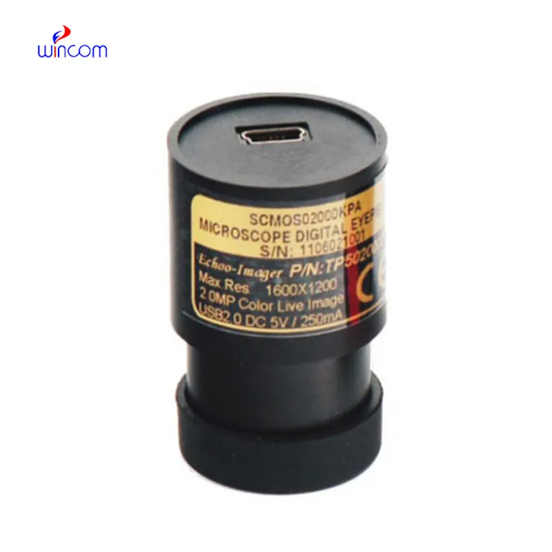
Q: What distinguishes a digital microscope from a traditional one? A: A digital microscope integrates cameras and imaging software, enabling users to view, capture, and analyze images directly on a computer or monitor. Q: How can vibration affect a microscope? A: Vibration can cause image blur or misalignment, so the microscope should always be placed on a stable, vibration-free surface. Q: What safety measures should be taken when using a microscope? A: Avoid touching optical parts with fingers, use slides carefully, and ensure electrical components are safely connected before operation. Q: Why is immersion oil used in some microscopes? A: Immersion oil increases the refractive index between the lens and specimen, improving resolution and brightness at higher magnifications. Q: How can you prevent mold growth in a microscope? A: Store the microscope in a low-humidity environment and use desiccants or dehumidifiers to keep optical components dry and mold-free.
The delivery bed is well-designed and reliable. Our staff finds it simple to operate, and patients feel comfortable using it.
This ultrasound scanner has truly improved our workflow. The image resolution and portability make it a great addition to our clinic.
To protect the privacy of our buyers, only public service email domains like Gmail, Yahoo, and MSN will be displayed. Additionally, only a limited portion of the inquiry content will be shown.
I’m looking to purchase several microscopes for a research lab. Please let me know the price list ...
Could you share the specifications and price for your hospital bed models? We’re looking for adjus...
E-mail: [email protected]
Tel: +86-731-84176622
+86-731-84136655
Address: Rm.1507,Xinsancheng Plaza. No.58, Renmin Road(E),Changsha,Hunan,China

 af
af
 es
es
 ar
ar
 tr
tr
 sw
sw
 pt
pt
 th
th
 ur
ur
 bn
bn
 ne
ne
 vi
vi
 km
km
 lo
lo
 de
de
 ru
ru
 fi
fi
 nl
nl
 fa
fa
 fr
fr
 ko
ko