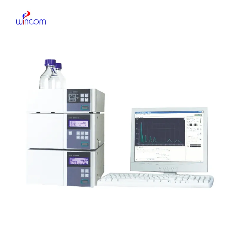
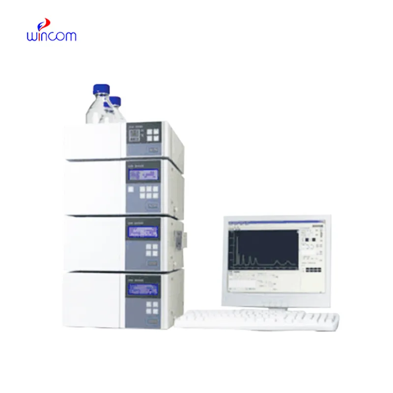
In medical and clinical laboratories, the use of size exclusion chromatography hplc results in highly precise determination of therapeutic compounds, metabolites, and biochemical markers. It facilitates creation of detailed patient sample profiles for research and diagnostics. The laboratory personnel prefer size exclusion chromatography hplc for confirming method reproducibility, validating analytical procedures, and keeping track of sample integrity. The ultrahigh sensitivity and versatility of the apparatus permit the laboratories to cater to varied applications, thus helping hospitals and research centers to provide reliable and accurate analytical results in various fields of science.
size exclusion chromatography hplc finds use in clinical toxicology laboratories to pinpoint and measure the amounts of possible poisons or drugs in abuse samples taken from patients. It is based on the separation of the various substances from complex mixtures like blood or urine, and that information is very important for the hospital doctors, who will then diagnose the case, decide on the treatment and monitor the patient’s safety.
In hospitals and clinical research, size exclusion chromatography hplc techniques will get higher resolution columns and ultrafast chromatography methods more and more. It will be possible to do these innovations in a shorter time and with a more accurate result. Future size exclusion chromatography hplc applications will be used to identify biomarkers quickly, monitor therapies in real-time, and manage patients more efficiently in both the laboratory and clinical settings.
Preventive maintenance is size exclusion chromatography hplc that play a very important role in clinical and hospital laboratories. The routine performance of flushing columns, cleaning injector valves, and monitoring pressure stability extends the life of the system. The laboratory staff is required to keep records of maintenance activities, replace consumables in a timely manner, and use solvents that are compatible. All of these practices are essential for the instruments' performance retention, lifespan extension, and high-quality analytical results, both in patient sample testing and research.
In today's laboratories, size exclusion chromatography hplc is indispensable for chemical analysis and serves as a primary instrument. Detection of compounds in intricate mixtures is first done through separation and then identification. Consequently, researchers can precisely check the interactions between molecules. size exclusion chromatography hplc is regarded to have extremely high reproducibility and it shares its strength with the fields of pharmaceuticals, biochemistry, and environmental science. Its alliance with sensitive detectors leads to the accurate measurement of very small amounts. size exclusion chromatography hplc is the trustworthy partner of lab technicians in validation of experiments, profiling of samples, and development of analytical methods. It not only gives consistent and detailed results but also boosts the efficiency of laboratories and at the same time, makes sure that the data obtained from research is reliable and thus, supports the advanced scientific inquiries that are conducted in various disciplines.
Q: What is HPLC used for in laboratories? A: HPLC turns out to be one of the most significant and essential analytical methods in laboratories equipped with the chemical compound analysis, separation, identification, and quantification of their presence in complex samples which are the research, clinical, and pharmaceutical applications. Q: How does HPLC separate compounds? A: The HPLC separation technique is based on the different affinities of the compounds to the stationary phase and mobile phase within the chromatography column. Q: Can HPLC analyze biological samples? A: Yes, it is certainly possible to carry out analyses on various biological fluids such as blood, serum, urine, etc. for the detection of metabolites, drugs, and biomarkers. Q: How often should HPLC columns be replaced? A: The replacement of the columns must be done according to the manufacturer instructions or when the performance begins to decline, which is quite usual after heavy use or contamination. Q: What detectors can be used with HPLC? A: The analysis type determines the use of, among others, UV, fluorescence, refractive index, and mass spectrometry detectors as the common detectors.
We’ve been using this mri machine for several months, and the image clarity is excellent. It’s reliable and easy for our team to operate.
This x-ray machine is reliable and easy to operate. Our technicians appreciate how quickly it processes scans, saving valuable time during busy patient hours.
To protect the privacy of our buyers, only public service email domains like Gmail, Yahoo, and MSN will be displayed. Additionally, only a limited portion of the inquiry content will be shown.
Could you share the specifications and price for your hospital bed models? We’re looking for adjus...
We are planning to upgrade our imaging department and would like more information on your mri machin...
E-mail: [email protected]
Tel: +86-731-84176622
+86-731-84136655
Address: Rm.1507,Xinsancheng Plaza. No.58, Renmin Road(E),Changsha,Hunan,China

 af
af
 es
es
 ar
ar
 tr
tr
 sw
sw
 pt
pt
 th
th
 ur
ur
 bn
bn
 ne
ne
 vi
vi
 km
km
 lo
lo
 de
de
 ru
ru
 fi
fi
 nl
nl
 fa
fa
 fr
fr
 ko
ko